Cause of ACNE SCAR
Acne scar cause can be summarized in a simple sentence. Acne scar is caused by improper and delayed treatment of inflammatory acne.
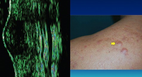
Acne scars are caused by healing of inflamed acne lesions. Skin repairs itself after any injury, including inflammation, by generating new collagen underneath. This deposition of new connective tissue causes acne scar . The main cause of acne scar is uncontrolled disease activity causing continuous inflammatory response within the dermis. The damaged skin repairs itself with scarring.
The acne scars form two pattern of scars:
1. Acne Scar with loss of tissue beneath the epidermis: These scars are depressed scars also known as atrophic scars and cause acne pits. These are caused by loss of support due to tissue damage in the dermis.
2. Acne Scar with increased tissue regeneration in the dermis: These are similar to hypertrophic scars and Keloids. They are typically raised, firm, reddish, and, at times, painful and itchy scars.
ACNE SCAR
Introduction
Scar is defined as “the fibrous tissue that replaces normal tissue destroyed by injury or disease.
Causes of acne scar – formation can be broadly categorizes as either the result or damage of “of increased tissue formation” or, more commonly ,loss of local tissue.
Acne scars are classified into three basic types – depending on width, depth and 3-dimensional architecture
Icepick scars – narrow (diameter <2mm), deep, sharply marginated and depressed tracks that extend vertically to the deep dermis or subcutaneous tissue
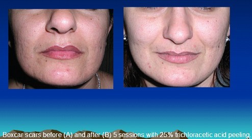
Boxcar scars – round ro oval depressions with sharply demarcated vertical edges. They are wider at the surface than icepick scars and do not taper to a point at the base. These scars may be shallow (0.1-0.5mm) or deep (≥0.5mm) and the diameter may vary from 1.5 to 4mm
Rolling scars – occur from dermal tethering of otherwise relatively normal-appearing skin and are usually wider than 4 to 5 mm in diameter. An abnormal fibrous anchoring of the dermis to the subcutis leads to superficial shadowing and to a rolling or undulating appearance of the overlying skin.
Other clinical entities included in this :
Hypertrophicscars – scar are raised within the limit of primary excision
Keloidal scars – scars transgress the boundary and may show prolonged and continuous growth
Sinus tracts may appear as grouped open comedones histologically showing a number of interconnecting keratinized channels.
Prevalence of acne scarring
One study reported acne scarring in 14% of women and 11% of men among 749 patients age between 25 and 58 years old.
Other publications suggest that between 30% and 95% of patients with acne develop some form of associate scarring.
Treatment option
Topical agents
Topical retinoids – Whether or not the use of topical retinoids improves acne scars that are already present has not been evaluated or quantified .
In an appropriately controlled study:
Topical retinoids such as tretinoin have been shown to increase dermal procollagen and collagen synthesis abd hence may provide some benefit in preventing scar development and potentially reduce the extent o scar formation that is in progress – unfixed scarring
There is no cogent evidence demonstrating that topical retinoids reduce scars that are already fully formed in the dermis – fixed scarring
Topical antimicrobial agents – Of no benefit on fixed scar
Topical corticosteroids – Intralesional triamcinolone injection for treatment of hypertrophic and feloidal scars is well established.
Long-term topical corticosteroid application is not recommended as local side effects such as atrophy and telangiectasia
Superficial Peeling
⦁ Very useful for treatment pigmented macular scars
⦁ Useful for improving boxcar scars
⦁ Improve active acne lesions
⦁ Can be utilized for dark skin
Superficial peelings include salicylic acid, 25% to 30% ; glycolic acid, 70% pyruvic acid, 40% and /or 50% 50 60%, trichloracetic acid, 20% to 30% and combination of salicylic acid or Jessner peel with trichloracetic acid.
Pathophysiology
Superficial peelings are utilized to induce a damage limited to the epidermis and papillary dermis. This results in epidermal regeneration and postinflammatory collagen neoformation.
Contraindications
⦁ Connective tissue disorders
⦁ Active skin disorders on the treatment sites
⦁ Oral anticoagulant treatment
⦁ Pregnancy
Photographic documentation – it is mandatory to obtain good-quality pictures before starting the procedure. This is an essential documentation for follow-up and for possible medicolegal issues.
Superficial peels are a cosmetic procedures that has the purpose of exfoliating the skin through the application of chemicals that induce skin irritation and damage
Expect severe burning during the procedure. This will usually last for 3 -4 minutes
Expect skin redness for 2 to 3 days
The skin will turn red brown and start to peel 2 to 3 days after peeling. Though rare, you can expect blisters and crusts.
The procedure can cause pigmented or white spots that are usually temporary and resolve in 1 to 3 months. In some skin types, however these pigmentary changes may persist and require specific treatments.
For the first week after procedure apply a moisturizer 3 to 4 times a day
Don’t scratch or remove the scales as it may result in scarring
Avoid sun exposure as it will cause development of pigmentary spots. Wear a high-protection sunscreen all the time for at least 2 months after procedure
Superficial peels improve the skin but may not completely eliminate acne scars
You may need to repeat the procedure 3 to 6 times for optimal results.
Medium depth and deep peeling
Agent include – Trichloroacetic acid (TCA), Croton oil, septisol, water, vegetable oils (glycerin, olive, sesame)
Dermabrasion for acne scars
Dermabrasion involves mechanically removing the epidermis and papillary dermis, creating a newly contoured open wound to heal by second intention
Reepithelialization of dermabraded skin occurs by upward migration of cells from the adnexal structures including hair follicles, sebaceous glands and sweat ducts
.jpg)
 Boxcar scars – round ro oval depressions with sharply demarcated vertical edges. They are wider at the surface than icepick scars and do not taper to a point at the base. These scars may be shallow (0.1-0.5mm) or deep (≥0.5mm) and the diameter may vary from 1.5 to 4mm
 Rolling scars – occur from dermal tethering of otherwise relatively normal-appearing skin and are usually wider than 4 to 5 mm in diameter. An abnormal fibrous anchoring of the dermis to the subcutis leads to superficial shadowing and to a rolling or undulating appearance of the overlying skin.
 Other clinical entities included in this :Hypertrophicscars – scar are raised within the limit of primary excision
 Keloidal scars – scars transgress the boundary and may show prolonged and continuous growth
 Sinus tracts may appear as grouped open comedones histologically showing a number of interconnecting keratinized channels.Prevalence of acne scarringOne study reported acne scarring in 14% of women and 11% of men among 749 patients age between 25 and 58 years old.Other publications suggest that between 30% and 95% of patients with acne develop some form of associate scarring.Treatment optionTopical agentsTopical retinoids – Whether or not the use of topical retinoids improves acne scars that are already present has not been evaluated or quantified .In an appropriately controlled study:Topical retinoids such as tretinoin have been shown to increase dermal procollagen and collagen synthesis abd hence may provide some benefit in preventing scar development and potentially reduce the extent o scar formation that is in progress – unfixed scarringThere is no cogent evidence demonstrating that topical retinoids reduce scars that are already fully formed in the dermis – fixed scarringTopical antimicrobial agents – Of no benefit on fixed scarTopical corticosteroids – Intralesional triamcinolone injection for treatment of hypertrophic and feloidal scars is well established.Long-term topical corticosteroid application is not recommended as local side effects such as atrophy and telangiectasiaSuperficial Peeling⦁ Very useful for treatment pigmented macular scars⦁ Useful for improving boxcar scars⦁ Improve active acne lesions⦁ Can be utilized for dark skinSuperficial peelings include salicylic acid, 25% to 30% ; glycolic acid, 70% pyruvic acid, 40% and /or 50% 50 60%, trichloracetic acid, 20% to 30% and combination of salicylic acid or Jessner peel with trichloracetic acid.PathophysiologySuperficial peelings are utilized to induce a damage limited to the epidermis and papillary dermis. This results in epidermal regeneration and postinflammatory collagen neoformation.Contraindications⦁ Connective tissue disorders⦁ Active skin disorders on the treatment sites⦁ Oral anticoagulant treatment⦁ PregnancyPhotographic documentation – it is mandatory to obtain good-quality pictures before starting the procedure. This is an essential documentation for follow-up and for possible medicolegal issues.Superficial peels are a cosmetic procedures that has the purpose of exfoliating the skin through the application of chemicals that induce skin irritation and damageExpect severe burning during the procedure. This will usually last for 3 -4 minutesExpect skin redness for 2 to 3 daysThe skin will turn red brown and start to peel 2 to 3 days after peeling. Though rare, you can expect blisters and crusts.The procedure can cause pigmented or white spots that are usually temporary and resolve in 1 to 3 months. In some skin types, however these pigmentary changes may persist and require specific treatments.For the first week after procedure apply a moisturizer 3 to 4 times a dayDon’t scratch or remove the scales as it may result in scarringAvoid sun exposure as it will cause development of pigmentary spots. Wear a high-protection sunscreen all the time for at least 2 months after procedureSuperficial peels improve the skin but may not completely eliminate acne scarsYou may need to repeat the procedure 3 to 6 times for optimal results.

 Medium depth and deep peelingAgent include – Trichloroacetic acid (TCA), Croton oil, septisol, water, vegetable oils (glycerin, olive, sesame)
 Dermabrasion for acne scarsDermabrasion involves mechanically removing the epidermis and papillary dermis, creating a newly contoured open wound to heal by second intentionReepithelialization of dermabraded skin occurs by upward migration of cells from the adnexal structures including hair follicles, sebaceous glands and sweat ducts


